• Link to X
  • Link to Facebook
  • Link to Youtube
Ecografía Deportiva
  • INICIO
  • FORMACIÓN
  • CASOS CLÍNICOS
    • Ver Todos
    • Autor
    • Articulaciones
    • País
  • ACTUALIDAD
  • ENTREVISTAS
  • PUBLICACIONES
  • F. JIMÉNEZ
  • Click to open the search input field Click to open the search input field Buscar
  • Menú Menú
Publicaciones & Investigaciones
Lesiones deportivas frecuentes
Artículos

Frequent Sports Injuries

Why the common muscle injury in athletes has become the Achilles heel for sports medicine doctors

GE and the NBA collaborate to provide doctors with clinical research to help treat common sports injuries

With the 2016 Olympics underway and team tryouts about to kick off for back-to-schoolers, late summer is the season of game time, not downtime. For elite or amateur athletes who are gearing up for competition, conditioning is key. But even the fittest among them are not without risk of injury.

MRI (side view) of an injured Achilles tendon.

MRI (side view) of an injured Achilles tendon.

From skyboxes to bleachers, fans watch the images on TV or direct from the stands – athletes in the midst of executing a critical play collapse, clutching a calf, quadricep or hamstring. It’s a sight startling enough to silence the most deafening sea of vuvuzelas. For the injured, forced to leave the game for medical attention, they await an answer, whether the injury is significant enough that it will prevent them from resuming competition.

Athletes and their fans have come to expect injuries as an unfortunate part of the game; muscle strains, tears, and pulls are the most common among them. Although most athletes have experienced some type of strain, sprain or pull, the technical term for these types of injuries is not widely known. A myotendinous injury impacts the areas where muscle transitions into tendon; it can impair performance, limit play time and can reoccur even after an athlete has healed and returned to play. Despite the prevalence of these types of injuries, very little research exists on how best to prevent and treat them. “These are basically soft-tissue injuries and these soft tissue injuries are common in all sports,” said Dr Fred Azar, chair of the research committee of the NBA Teams Physician Association. “Knowing the injury is not the issue but knowing the extent of the injury is why we need advanced research to help sort this out.”
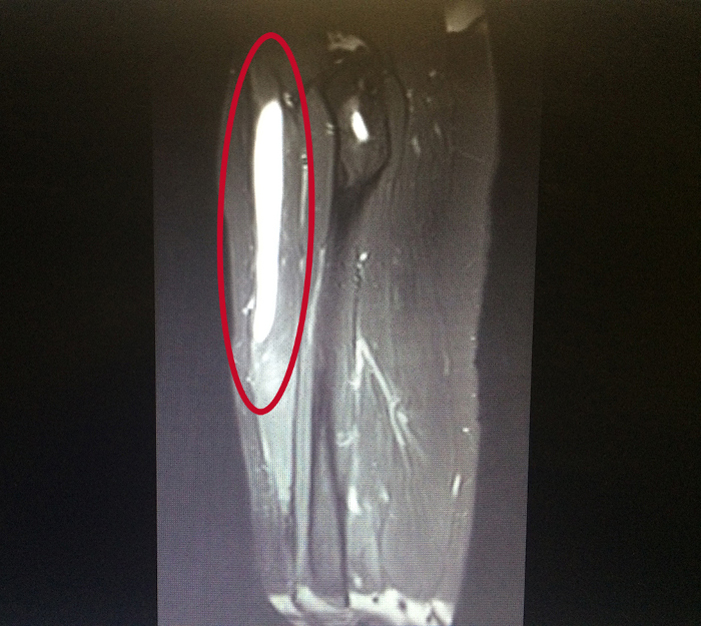
MRI (side view) of an injured quadriceps. The site of injury shows up bright white on the left of the scan.

MRI (side view) of an injured quadriceps. The site of injury shows up bright white on the left of the scan.

Typically, when athletes suffer injuries that affect the upper or lower extremities, doctors often utilize x-rays  and more advanced imaging technologies such as magnetic resonance imaging (MRI) or ultra-sound. Frequently occurring in the lower limbs, myotendinous injuries account for one third of all referrals to sports doctors.¹ Hamstring injuries are the most common form of these injuries in elite athletes who engage in sudden and rapid movements such as sprinting and jumping.² And although the frequency and debilitating impact of these injuries are well recognized, evidenced-based treatment options are not.

To better serve athletes and the doctors who treat them, in collaboration, the NBA and GE Healthcare are issuing a vUWAnyu_posts href=»https://gex.brightidea.com/MyotendinousCFP2016″>Call for Proposals (CFP) to researchers in orthopedics, sports medicine, radiology, and related disciplines asking for research and evidence-based treatment options that, up to this point, have been largely anecdotal. “We don’t know what we can tell people to do to avoid and best treat these injuries, and we’re hoping this research can answer that,” said Dr Kimberly Harmon, sports medicine physician at University of Washington and co-chair of the CFP along with Dr Azar. “We’ve been providing treatments like icing, massage and physical therapy for 40-50 years, and they seem to work but we really don’t know. It would be helpful if there was definitive research that looked at not only speeding return to play, but also prevention of recurrent injuries.”

MRI (side view) of an injured patellar tendon, which holds the kneecap in place.

MRI (side view) of an injured patellar tendon, which holds the kneecap in place.

By funding this program, the NBA and GE Healthcare are addressing high-priority clinical questions regarding the prevention, diagnosis, and treatment of acute and overuse injuries among NBA athletes, and importantly, to apply these findings to future basketball players, other athletes, and the general population. Dr. Azar believes in this myotendinous research collaboration between the NBA and GE Healthcare: “There is a lot of research out there but the answers are elusive and there’s no consensus. By knowing this information is scaled to other elite athletes, even the youngest athletes, we can use this information to help athletes in all sports, not just basketball.  It really transcends college, high school and professional sports to target the population for a broad reach.”

This is the second CFP sponsored by the NBA and GE Healthcare. The first vUWAnyu_posts href=»http://www.genewsroom.com/press-releases/nba-and-ge-healthcare-announce-first-round-research-winners-address-prevention?_sm_au_=i7VW3T5MNMwvRvnM»>round was dedicated to tendinopathy research.

The latest myotendinous CFP will award up to $1.5 million dollars over a three-year period to support preclinical and clinical research.  More information and complete CFP instructions and application forms are available at the NBA & GE Healthcare collaboration vUWAnyu_posts href=»https://gex.brightidea.com/MyotendinousCFP2016″>site.
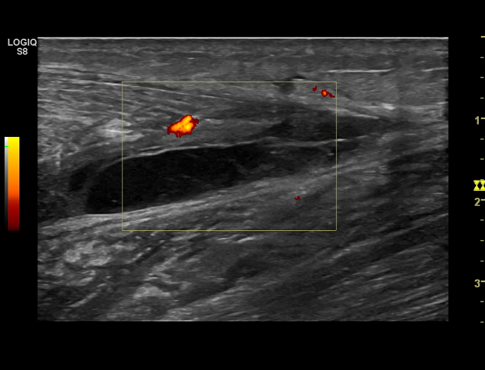
Ultrasound image: Ruptured calf muscle – image depicts a hematoma (blood clot and swelling within the tissues)

Ultrasound image: Ruptured calf muscle – image depicts a hematoma (blood clot and swelling within the tissues)

*Ultrasound photos provided by Dr. Fernando Jimenez, MD PhD, Universidad de Castilla la Mancha

¹ Krejci V, Koch P: Muscle and tendon injuries in athletes. Chicago, Yearbook Medical publishers, 1979

² Lieberman GM, Harwin SF: Pelvis, hip and thigh,: Sports medicine: principles of primary care. Mosby, 1997 pp 306-314

18 septiembre, 2017/por ecografiadeportiva.com
Compartir esta entrada
  • Compartir en Facebook
  • Compartir en X
  • Compartir en LinkedIn
  • Compartir por correo
https://ecografiadeportiva.com/wp-content/uploads/2017/09/Lesiones-deportivas-frecuentes.jpg 350 900 ecografiadeportiva.com http://ecografiadeportiva.com/wp-content/uploads/2016/11/logo-medical-sportoledo-200.fw_.png ecografiadeportiva.com2017-09-18 08:22:312017-09-18 08:29:09Frequent Sports Injuries
Artículos

Frequent Sports Injuries

Why the common muscle injury in athletes has become the Achilles heel for sports medicine doctors

GE and the NBA collaborate to provide doctors with clinical research to help treat common sports injuries

With the 2016 Olympics underway and team tryouts about to kick off for back-to-schoolers, late summer is the season of game time, not downtime. For elite or amateur athletes who are gearing up for competition, conditioning is key. But even the fittest among them are not without risk of injury.

MRI (side view) of an injured Achilles tendon.

MRI (side view) of an injured Achilles tendon.

From skyboxes to bleachers, fans watch the images on TV or direct from the stands – athletes in the midst of executing a critical play collapse, clutching a calf, quadricep or hamstring. It’s a sight startling enough to silence the most deafening sea of vuvuzelas. For the injured, forced to leave the game for medical attention, they await an answer, whether the injury is significant enough that it will prevent them from resuming competition.

Athletes and their fans have come to expect injuries as an unfortunate part of the game; muscle strains, tears, and pulls are the most common among them. Although most athletes have experienced some type of strain, sprain or pull, the technical term for these types of injuries is not widely known. A myotendinous injury impacts the areas where muscle transitions into tendon; it can impair performance, limit play time and can reoccur even after an athlete has healed and returned to play. Despite the prevalence of these types of injuries, very little research exists on how best to prevent and treat them. “These are basically soft-tissue injuries and these soft tissue injuries are common in all sports,” said Dr Fred Azar, chair of the research committee of the NBA Teams Physician Association. “Knowing the injury is not the issue but knowing the extent of the injury is why we need advanced research to help sort this out.”

MRI (side view) of an injured quadriceps. The site of injury shows up bright white on the left of the scan.

MRI (side view) of an injured quadriceps. The site of injury shows up bright white on the left of the scan.

Typically, when athletes suffer injuries that affect the upper or lower extremities, doctors often utilize x-rays  and more advanced imaging technologies such as magnetic resonance imaging (MRI) or ultra-sound. Frequently occurring in the lower limbs, myotendinous injuries account for one third of all referrals to sports doctors.¹ Hamstring injuries are the most common form of these injuries in elite athletes who engage in sudden and rapid movements such as sprinting and jumping.² And although the frequency and debilitating impact of these injuries are well recognized, evidenced-based treatment options are not.

To better serve athletes and the doctors who treat them, in collaboration, the NBA and GE Healthcare are issuing a vUWAnyu_posts href=»https://gex.brightidea.com/MyotendinousCFP2016″>Call for Proposals (CFP) to researchers in orthopedics, sports medicine, radiology, and related disciplines asking for research and evidence-based treatment options that, up to this point, have been largely anecdotal. “We don’t know what we can tell people to do to avoid and best treat these injuries, and we’re hoping this research can answer that,” said Dr Kimberly Harmon, sports medicine physician at University of Washington and co-chair of the CFP along with Dr Azar. “We’ve been providing treatments like icing, massage and physical therapy for 40-50 years, and they seem to work but we really don’t know. It would be helpful if there was definitive research that looked at not only speeding return to play, but also prevention of recurrent injuries.”

MRI (side view) of an injured patellar tendon, which holds the kneecap in place.

MRI (side view) of an injured patellar tendon, which holds the kneecap in place.

By funding this program, the NBA and GE Healthcare are addressing high-priority clinical questions regarding the prevention, diagnosis, and treatment of acute and overuse injuries among NBA athletes, and importantly, to apply these findings to future basketball players, other athletes, and the general population. Dr. Azar believes in this myotendinous research collaboration between the NBA and GE Healthcare: “There is a lot of research out there but the answers are elusive and there’s no consensus. By knowing this information is scaled to other elite athletes, even the youngest athletes, we can use this information to help athletes in all sports, not just basketball.  It really transcends college, high school and professional sports to target the population for a broad reach.”

This is the second CFP sponsored by the NBA and GE Healthcare. The first vUWAnyu_posts href=»http://www.genewsroom.com/press-releases/nba-and-ge-healthcare-announce-first-round-research-winners-address-prevention?_sm_au_=i7VW3T5MNMwvRvnM»>round was dedicated to tendinopathy research.

The latest myotendinous CFP will award up to $1.5 million dollars over a three-year period to support preclinical and clinical research.  More information and complete CFP instructions and application forms are available at the NBA & GE Healthcare collaboration vUWAnyu_posts href=»https://gex.brightidea.com/MyotendinousCFP2016″>site.

Ultrasound image: Ruptured calf muscle – image depicts a hematoma (blood clot and swelling within the tissues)

Ultrasound image: Ruptured calf muscle – image depicts a hematoma (blood clot and swelling within the tissues)

*Ultrasound photos provided by Dr. Fernando Jimenez, MD PhD, Universidad de Castilla la Mancha

¹ Krejci V, Koch P: Muscle and tendon injuries in athletes. Chicago, Yearbook Medical publishers, 1979

² Lieberman GM, Harwin SF: Pelvis, hip and thigh,: Sports medicine: principles of primary care. Mosby, 1997 pp 306-314

18 septiembre, 2017/por ecografiadeportiva.com
Compartir esta entrada
  • Compartir en Facebook
  • Compartir en X
  • Compartir en LinkedIn
  • Compartir por correo
https://ecografiadeportiva.com/wp-content/uploads/2017/09/Lesiones-deportivas-frecuentes.jpg 350 900 ecografiadeportiva.com http://ecografiadeportiva.com/wp-content/uploads/2016/11/logo-medical-sportoledo-200.fw_.png ecografiadeportiva.com2017-09-18 08:22:312017-09-26 10:37:09Frequent Sports Injuries

Search

Search Search

CASOS CLÍNICOS

  • POR ARTICULACIÓN
    • Hombro
    • Codo
    • Cadera
    • Mano y Muñeca
    • Rodilla
    • Pié y Tobillo
    • Otras Lesiones
    • Intervencionismo
  • POR AUTOR
    • Dr. Adolfo E. Reque
    • Dr. Alfonso Martínez Franco
    • Dr. Américo Enrique Villalobos
    • Dr. Ankit Shah
    • Dra. Begoña Manuz
    • Dr. Bruno Sagástegui
    • Dr. César León
    • Dr. Eduard Ribas i Forcadell
    • Dr. Felipe Giraldo
    • Dr. Fernando Jiménez Díaz
    • Dr. Francisco Lara
    • Dr. Germán Arancibia
    • Dr. Gustavo Reque
    • Dr. Iván J. Ardines
    • Dr. Iván Quirós
    • Dr. Javier Ferreira Villanova
    • Dr. Jose G. Villa
    • Dr. Jose Mª Bueno
    • Dr. Jose Mª Urraca
    • Dr. Marcelo A. Ortiz Obregón
    • Dr. Maulik S. Patel
    • Dr. Nazul Aké
    • Dr. Pablo dos Santos
    • Dr. Philippe Moulin
    • Dr. Pramod Bapna
    • Dr. Rafael Romero
    • Dr. Ricardo Uribe Gonzalez
    • Dr. Ricardo Díaz
    • Dr. Ronal Huapaya
    • Dr. Samuel Pina
    • Dr. Satish Pawar
  • POR PAIS
    • Chile
    • Colombia
    • Emiratos Árabes Unificados
    • España
    • Francia
    • México
    • Perú
    • República de La India
    • Uruguay

Últimos casos…

MEDICAL SPORTOLEDO

Calle Oslo, 3 – 1º izq.
TOLEDO

Tlf.: (+34) 925 210 757
ecografiadeportiva@ecografiadeportiva.com
CONTACTO

PUBLICACIONES RECIENTES

Consulta los últimos libros publicados por el Dr. Fernando Jiménez Díaz

SITIOS DE REFERENCIA

Diferentes Centros de referencia y Sociedades médicas relacionados con la medicina deportiva
X Logo X Logo Seguiron X RSS Feed Logo RSS Feed Logo Suscribirsea canal RSS
© Copyright - Medical Sportoledo
  • Link to X
  • Link to Facebook
  • Link to Youtube
Link to: Fractura de peroné  Weber B. Link to: Fractura de peroné  Weber B. Fractura de peroné  Weber B. Link to: Frequent Sports Injuries Link to: Frequent Sports Injuries Frequent Sports Injuries
Desplazarse hacia arriba Desplazarse hacia arriba Desplazarse hacia arriba